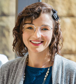
Epilepsy patient gives back to science

After life-changing medical care, patients often want to give back. For Jasmine Williams, that opportunity came before brain surgery to cure her epilepsy.
Determined to gain control of her seizures, Jasmine Williams had brain surgery at UCI Medical Center in June 2018. It turned out to be life-changing — for herself and potentially many other people with neurological conditions.
Now seizure-free, Williams, 28, is able to hold her first full-time job: teaching fifth grade in Montclair, Calif. She’s able to drive, so she no longer relies on her family to get places.
She’s also pursuing her passion for woodworking, brightening the lives of children with serious illnesses by donating handcrafted jewelry cases to Beads of Courage, an organization that gives pediatric patients a bead for each medical test they undergo.
For the good of science
Williams already has helped advance brain science by participating in more than two dozen research trials while undergoing presurgical tests to pinpoint the origins of her seizures.
Data from her case has contributed to groundbreaking published studies that are shedding light on neurological disorders, sleep and how humans learn.
“I think a patient who has epilepsy understands other people who are suffering,” says Dr. Jack J. Lin, director of the UCI Health Comprehensive Epilepsy Program and professor of neurology and biomedical engineering at UCI.
“It seems like there’s a special quality that leads to this kind of generosity.”
More than 100 seizures a night
Williams was referred to Lin after years of suffering from nightly seizures — more than 100 some nights — and the dizzying side effects of epilepsy medications.
“Dr. Lin gave me so much hope,” says Williams, who had her first seizure at age 8. “He explained what the surgical process would be like, he took his time with me and he was honest with me.”
Surgery can be an excellent option for eligible patients who struggle to control epilepsy with medications.
“The success rate is really high,” says Dr. Frank P.K. Hsu, professor and chair of the Department of Neurological Surgery at UCI School of Medicine and professor of biomedical engineering at UCI.
“About 60% to 70% of patients will have no more seizures. That’s great, but if you can reduce the frequency and severity of seizures that, too, can make a big difference in the patient’s life.”
Mapping electrical activity
Before surgery, Williams underwent two multiday evaluations to determine where her seizures were originating in her brain. Hsu made 18 small holes in Williams’ skull, feeding wires into the holes to insert electrodes in her frontal and temporal lobes.
Data from these tests allowed Williams’ medical team to gather critical details about her case — confirming that her seizures were originating in the frontal lobe.
UCI researchers compiled additional data for more than a dozen clinical studies by recording her reactions on various tests.
“This information is so precious because there’s no other way of getting it,” says Lin. “We are indebted to our volunteer patients in our Comprehensive Epilepsy Program.”
Inside Williams’ brain
Some of the studies asked Williams to view images on a screen and answer questions; others involved recording her brain activity as she slept.
“It was actually kind of fun,” she says. “I was happy to just give back.”
Because Williams had electrodes in her amygdala region (the brain’s emotion center) as well as in her hippocampus region (the memory center), she also participated in a study — recently published in the journal Neuron — about how the amygdala and hippocampus communicate with one another.
“We are discovering how emotional memory occurs inside a human brain in real time,” says Lin. “This will have profound implications for conditions such as post-traumatic stress disorder.”
Pinpointing her seizures
In another procedure called subdural electrode grid testing, Hsu arranged electrodes in a section of Williams’ frontal lobe.
“This grid allows us to map exactly the extent of the seizure location,” says Lin.
“It also allows us to map the functional activity around the area. We send a little electrical activity into these 3-millimeter contacts and we see if she’ll have any behavior change, like a twitch of an arm, finger or mouth.”
This testing is critical to ensure that brain tissue affecting motor function is spared during surgery.
Williams’ procedure involved removing a few centimeters of brain tissue — about the size of a kiwi, says Hsu — and implanting a responsive neurostimulation device as insurance to prevent future seizures.
She was cleared to go home the following day and has been seizure-free ever since.
“In the end, I couldn’t have hoped for a better result,” Williams says.


